This article on Epainassist.com has been reviewed by a aesculapian professional , as well as checked for facts , to assure the readers the best potential truth .
We follow a nonindulgent editorial policy and we have a zero - tolerance insurance policy regarding any level of plagiarization . Our article are resourced from reputable on-line pages . This article may bear scientific references . The numbers in the excursus ( 1 , 2 , 3 ) are clickable links to peer - reviewed scientific papers .
The feedback link “ Was this clause Helpful ” on this page can be used to report content that is not exact , up - to - date or questionable in any manner .
This clause does not supply medical advice .
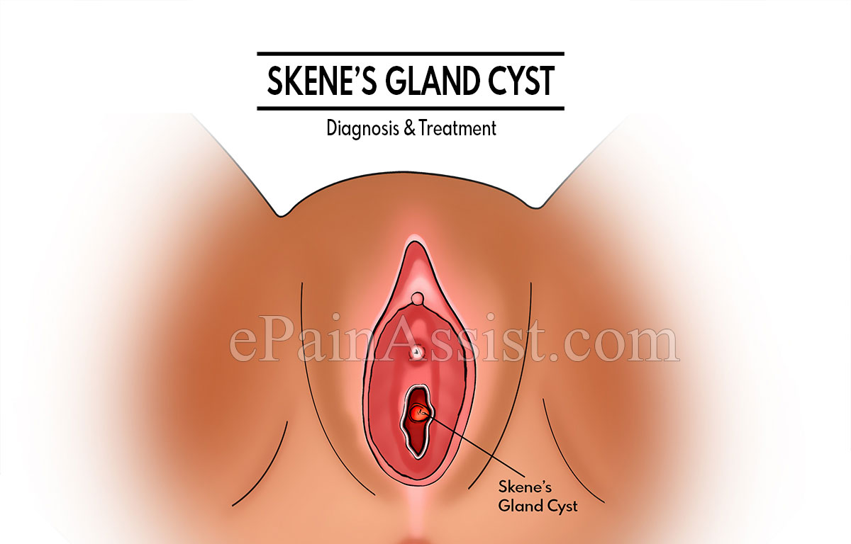
Skene ’s glands , also known as less vestibular glands are locate on either side of the urethra in womanhood . While the precise role of the gland is not full see , it is believed to be concern to female interjection during intimate rousing . In some rarefied cases , askene ’s gland cystmay be mention , possibly due to occlusion of the glands .
Skene ’s glands are also roll in the hay as paraurethral secretor and are bilateral prostate homologues glands . It was first discovered and identify by Alexander Johnston Chalmers Skene in 1880 . The impedimenta of Skene ’s ducts due to infection , inflammation due to skenitis due to gonorrhea or cystic degeneration of embryonic remnants of the paraurethral glands can cause paraurethral cyst . This condition is called Skene ’s gland vesicle , which is often rare.1
Skene ’s glands are periurethral or paraurethral glands located adjacent to the distal urethra . The tissue paper surrounding them are a part of the clitoris . These glands secrete a pith to lube the urethra opening . The kernel is cerebrate to be anti - microbic and can prevent urinary tract infections . Although the exact role of these secretory organ is not clear , it is believed to be relate to female ejaculation during intimate arousal.2
What is Skene’s Gland Cyst?
Skene ’s secretor vesicle is considered rare , however , some sheath have been report affecting various age groups . Skene ’s canal cyst develops near the distal urethra and can cause perineal discharge , dyspareunia , or abscess formation in some case .
Most people with Skene ’s secreter vesicle may not have any symptom or some experience minor irritation during urination or sexual activity .
It is believed that Skene ’s gland cyst is holding cyst that result from secondary to instigative obstruction of the Skene ’s ducts in females.3 Most Skene epithelial duct cysts are less than 1 cm and ordinarily do not cause any symptoms , while some larger 1 can make complaint and evening cause dyspareunia .
Skene ’s secretor vesicle form when the duct gets obstructed either due to contagion or inflaming . It can get septic lead inurinary tract infectionsand abscesses . Some large Skene ’s secretory organ cyst or cyst in the epithelial duct can even obstruct the urethra and restrict the current of pee . This can get trouble in passing urine and can also cause pain in the urethra . The symptom mark in such case admit hesitating start at the first of urination , dribbling at the end of urination , and keeping of piddle . Large vesicle can make pain during urination and even during sexual intercourse .
In very rare cases , abscess may be formed due to the vesicle , which can be tender , very painful , and get egotistic . The tegument over the surface area may come out red . Fever is normally not to be present . It is either treat with antibiotics or may have to be removed surgically.4
How are Skene’s Gland Cysts Diagnosed and Treated?
Skene ’s gland cyst is usually diagnosed on clinical examen . It is usually done when the vesicle is symptomatic and aesculapian help is attempt . In most cases , the diagnostic cyst are tangible and can be diagnosed clinically . Sometimes , asymptomaticcystsmay also be noted during pelvic examination for other purpose . During a pelvic scrutiny , the doctors may feel the secretor cyst or abscess , if they are large .
However , in some case , for better viewing or confirmatory diagnosis additional tryout may be done . Usually , ultrasonography or cystoscopy ( using a flexible tube to view the bladder ) may be done to confirm the diagnosis . In some cases , if needed , additional scan like MRI may be done . It may appear as round or oval Mass located laterally to the external urethral meatus and deficient to the pubic symphysis.3
Other conditions that must be considered before making a diagnosis include the Bartholin gland vesicle and urethral diverticulum or urethrocele .
Treatment of Skene ’s secretor cyst mainly depends on the presence and severity of the symptom and the size of the cyst .
What Do Some Case Reports Of Skene’s Gland Cyst Say?
While it may be noted in adults , type have been reported when females of varying ages have abide from Skene ’s secretory organ cyst . A study report about a case of Skene ’s secretory organ cyst in a 1 - day - old female infant states that it is a rare congenital anomaly in female newborns baby . Although forcible examination alone can help diagnose the condition , get laid about the condition can help oneself plan only necessary investigation and interventions.5
Skene ’s secreter secrete a mucoidal fabric with intimate stimulation . However , in newborns , the paraurethral glands may sometimes answer to maternal estrogen and secrete the mucoidal substance . The condition is rare , especially in newborn and the precise cause is not known . But mayhap conditions like an infection , inflammation , or cystic degeneration of embryonic oddment of the paraurethral secretor can lead to the obstruction of Skene ’s ducts .
In newborn , the condition may present as an symptomless bulging mass with small vessel on the Earth’s surface , located on either side of theurethralmeatus . Skene ’s gland cyst may have no reflection , but a careful study of the urinary tract is necessary to exclude tortuousness or dissimilar severe injuries.6
Here is about another case of Skene ’s glands cyst in a 37 - twelvemonth - old charwoman . She presented with a charge of external dyspareunia . There were no urinary symptoms and the physician did not notice any obstructer of the urinary catamenia . History taking revealed that the mass had been growing for almost 8 years and was never painful . Pelvic and rectal examination were normal and there was no extrusion of pus or urine from the urethral meatus on giving pressure on the mass.7
References :
