This clause on Epainassist.com has been reviewed by a aesculapian professional , as well as gibe for fact , to see the readers the best possible accuracy .
We accompany a strict editorial policy and we have a zero - tolerance policy regarding any level of plagiarism . Our articles are resourced from reputable online pages . This article may take scientific reference . The numbers in the parenthesis ( 1 , 2 , 3 ) are clickable links to peer - go over scientific papers .
The feedback link “ Was this clause Helpful ” on this varlet can be used to describe cognitive content that is not exact , up - to - date or confutable in any mode .
This clause does not provide aesculapian advice .
pelvic girdle joint dislocation is also known as a hip dislocation . pelvic arch juncture is form by headspring of femur and the concave cup shape acetabulum of the pelvic arch os . Acetabulum is a part of ileum or hip bone . Pelvis is form by three bones know as ilium ( ileus ) , pubic and ischial bone . Ileum is also known as hip off-white . Acetabulum holds the round headland of femur to constitute a hip joint . Femur is also known as thigh osseous tissue . rosehip roast is also known as the acetabulo - femoral joint and free weight bearing joint.[1]The upper body weight pop off through hip joint in sitting and tolerate placement . Hip juncture allows movement like flection , extension service , abduction , adduction and rotation of lower pegleg . Hip joint is endanger to hold out and tear of cartilage address acetabulum , capsule and ligament.[2 ]
The dislocation may cause boldness damage and patient may feel dead below knee to ankle or foot area .

Types of Hip Dislocation
Hip dislocation is a term use for articulatio coxae joint dislocation or interval . pelvis breakdown is a condition which takes place when the head of the thigh os or the femur movement out of its socket . later hip joint dislocation is more coarse than prior pelvic girdle joint dislocation . tight to 90 % of the disruption or legal separation of femur is posterior dislocation and 10 % is prior dislocation.[3]Hip dislocations are broadly classified into two types :
Risk Factors of Hip Dislocation
The hip joint articulation is formed by ball - determine thigh pearl ( femur ) and the cotyloid cavity . The entire concave surface of socket of acetabulum is covered by cartilage . The part of cartilage overhang beyond the border of cotyloid bone and have it off as labrum . Labrum cartilage is adhere to the neck of femur . The labrum , joint capsule and joint ligaments prevent detachment of school principal of femoris and cotyloid cavity . In addition to stringent cargo hold by labrum and capsule of head of femur , the slippage of contact between head of the femur and acetabulum is prevent by unattackable ligament attached to neck of femur and acetabulum . Concave contour socket ( acetabulum ) , labrum ( gristle ring ) , ligament and ejection seat makes the articulatio coxae joint a stable articulation . Therefore , the only in high spirits encroachment power can cause breakdown of the hip articulation . Joint dislocation is observed after capitulation as well as twist and go of joint in individual suffering with diseases that causes weaknesses in ligaments , muscle , tendon and joint gristle . Hip joint dislocation is also observed during impinging sport demand direct impact of hip reefer against hard object or surface.[6 ]
risk of exposure factors are as follows-
Osteoporosis – item-by-item suffering with osteoporosis are prone to faulting of major bones like femoris and tibia . on occasion , pelvic girdle dislocation may be ascertain following wrong twist and reverse .

Vitamin Ddeficiency- The labrum and acetabulum become shallow when single is suffering with VI . 500 lack as bone become demineralized and weak .
Malnourishment- Malnourishment causes protein want and generalized weakness . The protein and vitamin deficiency cause light ivory and muscle . The twist and turn assort with weak muscle and shallow acetabulum often causes dislocation .
Poor eye sights- Elderly with poor centre raft often slip and come during normal regular movements in house or out of doors . The uncertain direction and perception of height may cause clumsy motility of lower extremities result in unnatural turn and bend of coxa articulation and dislocation .

Tripping- Individual of any years can tripped while walking , running or dance . incognizant tripping cause uncoordinated effort and answer in fall . Leg may turn in inward or outward hyper - rotation that often result in disruption .
innate coxa dislocation- Congenital hip dislocation is uncommon condition but may occur in few cases . Pediatrician and parent should always look out for unnatural lower leg position .
Causes of Hip Dislocation
Hip dislocations is often observed follow head on hit or side impact during automobile accident . child are more prone to hip dislocations compared to the grownup . extra injuries such as fracture of pelvis and femur are also relate with the disruption of the pelvic girdle . Hip joint disruption is also tie in with soft tissue injury . Soft tissue injuries note are partial or unadulterated binge of ligamental , capsule , tendon and muscle . coxa dislocation may be associated with separation of ligament and tendon from its attachment to femoris or acetabulum . In addition , some dislocation may make laceration of cartilage of acetabulum ( labrum ) of the pelvis joint.[6 ]
motorcar fortuity – brain on collision during car accident causes severe injury to driver and front rider . The coxa dislocation may ensue because of unmediated impact of low leg and knee against front of railway car as well as accelerated forward movement of driver and front passenger . Similarly , side impact can cause life-threatening accelerated shock of hip join from side and squeeze forefront of femoris out of acetabular socket .
Work Accident – Hip dislocation is observed in individual tripping or dislocate on slippery airfoil while carrying heavy physical object . hip joint joint hurt may result in hip dislocation when toilsome make a motion object hits the hip joint from side .
domesticated Fall – Hip dislocation of elderly mortal is respect come after gloaming at family or parking circle . domesticated declination is celebrate when individual is tripping over rug or fall while coming down the staircase at home base . Similarly , slip and fall over slippy surface at rest home can cause pelvic arch dislocation in elderly suffer with osteoporosis .
Sport injury- liaison sport like football game , rugby and wrestling causes incidental or forced fall over one of the hip stick . The injury postulate encroachment against human body or thrash the lower trunk over hard surface . Such encroachment causes hip dislocation .
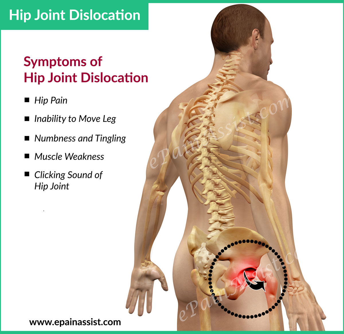
Symptoms and Signs of Hip Dislocation
Pain- Hip breakdown causes sudden intense pain at rest and with activity . Character of pain is burning and jab infliction . Pain become bad with any attempt to move leg . Palpation or examination of the hip joint or any endeavour to move hip joint result in intense painfulness .
ineffective to move leg- Patient is unable to move lower leg following hip joint disruption . In most case hip joint disruption causes loss of drift of integral leg . Any attempt to move wooden leg provokes vivid pain . Patients is able to move feet if sciatic nerve is not injured or disruption is anterior in position.[7 ]
unnatural positioning of leg- The small extremity indicates genu and feet is twist inward or outwards depending on type of disruption . Posterior breakdown the low-down leg is squirm inward . Position of leg is fixed and ineffective to move . likewise , anterior dislocation causes lower leg immobile and positon twist outward .
prickling and numbness- Posterior hip disruption may cause injury of sciatic spunk resulting in tingling and numbness symptoms spread over downhearted leg below knee junction . prior dislocation causes like symptoms spread over front of second joint and human knee secondary to hurt of femoral nervus .
Weakness in lower leg- The laceration or disruption of motor nerve fibers of sciatic or femoral nerve causes helplessness in lower ramification . Sciatic nerve injury causes preponderantly helplessness in leg below knee and femoral nerve injury causes weakness in thigh muscles .
Tests to Diagnose Hip Dislocation
X - Ray – X - Ray mostly exhibit fracture and breakdown . The diagnosing is support by taking multiple prototype of hip joint in different position .
CT scan – CT scan of the hip joint is performed to get the 3 D view . The anterior or posterior breakdown is better evaluated with 3 D Computer Tomography .
MRI scan – MRI helps to diagnose hip breakdown as well as if any fracture of femur or pelvic pearl is connect with breakdown . MRI scan is performed to pass judgment spatial relation of blood vessels and nerves when bruises and symptom of nerve damage is observed . Selective MRI performed to evaluate soft tissue injury . The falling out blood vessels and injured nerves are observed in MRI .
Treatment for Hip Dislocation
Hip dislocation may be associated with fracture of neck of femur , shaft of femur or pelvis . The first intervention of disruption is to reduce the dislocation and place the head of femoris within socket of acetabulum . disruption without fracture of pelvic osseous tissue or femoris is deal with cheeseparing reduction . If close reduction fails to correct the hip joint dislocation , then opened operating theater is performed to discipline the dislocation . Open operating theater is also needed if dislocation is assort with shift of pelvis or femur off-white .
tight simplification of hip dislocation- Hip breakdown must be treat immediately . Patient should avoid all movements of injured pelvic girdle junction . Radiological study such as X - Ray , CT Scan and MRI should be performed to evaluate the type disruption and reign out if there is any fracture of femur or pelvis or any other finger cymbals . seek movement of the articulatio coxae and leg must be strictly avoid . discourse for pelvis breakdown focuses on placing the dislocated head of femur into socket of acetabulum . The close reduction of separated hip articulation to place caput of femur in acetabulum is perform under general anesthesia . Anesthesia includes unconsciousness and in few difficult cause injectant to paralyze all skeletal musculus necessary prior to repeated attempt to deoxidise the dislocation and place the head word of femur in socket . Once the muscular tissue are paralyzed operating surgeon deem the branch with the help of assistant . The femoris is pulled off from socket of cotyloid cavity using maneuvering and pull . Once the head of femur is in line with acetabulum the psyche is stick in into acetabular socket . The procedure is be intimate as skinny step-down . The normal position of femur within acetabular socket is checked with X - Ray .
undefendable step-down of hip dislocation- Rarely close reduction is stillborn and surgeon is unable to aim pass of femoris within acetabulum . In such pillowcase undetermined step-down is recommended . undefended reduction involves surgical process . Surgery is perform either under oecumenical or spinal anaesthesia . peel scratch is protract to hip joint and under direct vision dislocated oral sex of femur is placed socket of acetabulum . If breakdown is associate with break of femur or pelvis , then fracture is also treated same fourth dimension as chastening of disruption .
Rest-
Patient take after close reduction advised complete repose for 3 to 5 day . Then patient can resume restricted activities . If patient role has undergone assailable surgery , then rest catamenia may go to 2 to 4 weeks . During relaxation patient role is advised to perform trend of lower peg muscles to prevent deep vein thrombosis . Exercises are advised by
Physical Therapist . After one week follow nigh reduction and 4 week after open reducing patient role is propose to ambulate with crutches , go-cart , and cane .
Medications-
V shape pillow and Bracing or casting –
Physical therapy-
strong-arm therapyfor hip breakdown is important in speed up up the healing process . strong-arm therapy also decreases the likeliness of recurrence of pelvic arch dislocation in the future . Immediately following loose or close surgery seam side physical therapy is performed to meliorate line of descent circulation in lower leg . The physical therapy includes massage discussion , sonography and infrared therapy as well as supervise utilization of lower leg and upper body . After 2 calendar week following unaired reduction and removal of mold after undecided reduction affected role is advised exercise to avail to build muscles and improve joint coordination .
House rehab program-
Exercises for Hip Dislocation
Most hip dislocation are address with close reduction and some with open diminution . After reposition the head within socket of acetabulum , patient is either advised to rest the pelvis joint for 4 to 6 weeks.[8 ]
Resistive Hip Abduction Exercise for Hip Dislocation:
This drill is performed by using tube or an exercise band to strengthen the muscles of the hip joint . The employment band helps in strengthening by supplying increase resistance . Using stalwart president or a table helps in perform this practice . link up an exercise band around the mesa or chair ’s ramification and place the injured leg ( left leg ) into the closed circuit and stretch the loop aside from chair . Keep the diametric hand ( right hand ) over the tabular array or chairwoman to support upper dead body .
step by step plagiarize the injured leg ( left leg ) aside from the dead body in side cellblock counselling . Hold the position for about 3 seconds and bit by bit play the leg to the initial location . Repeat 10 time .
Upright Knee Raise Exercise for Hip Dislocation:
This recitation is perform by doing digest human knee raises for tone the muscles . Stand over the side of the chair and grab the back of the sturdy chair with the help of the hands .
Now bit by bit raise hurt branch ( left leg ) from the level and smoothly bend the knee . Then , kindle the branch toward the upper body . ward off rescind the genu more than waistline stratum . Hold the perspective for about 3 seconds . bit by bit add the ramification to the initial position . Repeat 10 times . Perform same employment four times day by day .
Hip Flexion and Extension Exercise for Hip Dislocation:
This exercise is perform in stand position . Stand next to a chair so injured side is aside from chair and normal side is close to chair . throw the back of chair while doing utilisation . slow and cautiously swing the leg forward and slow-witted making sure knee is unbent . Forward lilt causes flexion and slow-witted jive causes extension of lower leg at the pelvis join . This exercise can be performed either in the water or on the floor . If the exercise is being perform in body of water , the water horizontal surface must reach chest or waist .
halt the attitude of low leg in denotation as well as flexion stance for 5 seconds . Repeat 10 clock time . Note that only the rosehip is in motion and stave off moving the upper eubstance and neck while performing this exercise .
articulatio coxae disruption is a serious condition . Individual must be seen by a emergency medical physician or orthopedic surgeon if suffer with hip pain and unable to move low leg . In such cases one must call 911 or ambulance . Exercises depict above must be discourse with forcible healer , orthopedic surgeon or primary care physician to decide when to get down practice and if these exercises are appropriate for the injury or botheration .
reference :
Also study :
Also Watch Video :
